 You are likely familiar with what an X-ray looks like, even if you’ve never had one before. Whether you’ve seen them on TV or in movies, you will recognize X-rays for their distinct black, white, and gray images of the bones in the body. X-rays are common in medical settings, especially for diagnosing injuries like a broken bone or dislocated joint. You might be looking to find Snellville imaging that offer X-rays near you. Many places offer X-rays, including hospitals and urgent care centers. If you have a musculoskeletal injury like a broken bone or joint injury, then consider going to see an orthopedic doctor for an X-ray and quality, comprehensive care for your injury or issue.
How X-Rays Work
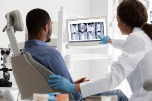
X-rays are a fast and reliable way for doctors to diagnose issues and injuries that affect bones and joints. Updated and improved X-ray technologies now mean that an X-ray will be an electronic image that your doctor can see almost instantly after the scan. Depending on where you have the injury or issue, an X-ray will scan that area and send a small amount of radiation through that part of the body. Bones are made of calcium, and radiation cannot pass through them, so they will show up as white or lighter shades of gray on the scan. For example, the rib bones will show up as white and lighter gray, while the lungs will be dark gray. A radiographer operates the X-ray examination and then sends the scan results to a radiologist for interpretation.
What X-Rays Are Used For
X-rays are used for diagnostic purposes in a number of injuries and conditions. Doctors rely on X-rays due to their fast and reliable results, especially in emergency or time-sensitive settings. X-rays are typically recommended for diagnosing musculoskeletal conditions because they will provide doctors with a quick and clear image of any damage to bones or joints. However, unlike CT scans or MRI scans, X-rays do not provide doctors with imaging of muscles, tendons, or other soft tissues in the body. With injuries or issues that may also affect soft tissues, your doctor may first request an X-ray to rule out any potential bone or joint-related injuries before moving on to other types of diagnostic imaging tools.
Common Injuries X-Rays Can Detect
Here are five examples of what X-rays can detect and how a doctor uses these for diagnosis and treatment. Snellville imaging experts can provide further direction and treatment with all of these diagnoses.
Broken Bones
The most common reason for X-rays is to detect broken bones. A broken bone is also known as a fracture and can occur in a few different ways. When many people think of a broken bone, they picture a brightly colored cast on a broken arm or leg. However, a broken bone doesn’t always need a cast, and in some parts of the body, it isn’t even possible. There are also types of broken bones known as hairline fractures, where a cast may not be beneficial.
Joint Injuries
Another reason a doctor may request an X-ray is for a joint injury. If you twisted your ankle walking down some steps or tripped and caught yourself with your hands outstretched, then your doctor may want to use an X-ray to check for any joint damage in your ankle joint, wrist joint, or elbow joint. An X-ray will show whether or not any bones have been broken or whether the joint is out of place, also known as a joint dislocation.
In addition to identifying fractures near a joint, X-rays can also reveal subtle alignment issues that may not be obvious during a physical exam. For example, even a partial dislocation, known as a subluxation, can sometimes be detected through imaging. X-rays may also show joint space narrowing, which can indicate cartilage loss or degeneration following an injury. In traumatic accidents, physicians often use X-rays to ensure that joint surfaces remain properly aligned and stable. Detecting these issues early is important, as untreated joint injuries can lead to chronic instability, stiffness, reduced range of motion, or long-term complications such as post-traumatic arthritis.
Bone Conditions
Doctors utilize X-rays to diagnose some medical conditions that affect the bones, like arthritis or osteoporosis. Images from an X-ray scan will show whether or not your bones have been damaged by one of these conditions or if there are any abnormalities that require further intervention.
In addition to diagnosing arthritis or osteoporosis, X-rays can also help doctors identify bone infections, structural deformities, or even metabolic bone disorders. Certain conditions may cause bones to appear thinner, denser, or irregularly shaped on imaging. For example, bone spurs, which are small bony projections that develop along the joint edge, can often be visible on X-rays and may also cause pain or limited movement. In children and adolescents, X-rays can evaluate growth plate development and detect abnormalities that could affect long-term bone growth. By identifying these changes early, doctors can recommend treatments that help slow progression and preserve overall bone health.
Cancers
Doctors also use X-ray technologies to diagnose certain cancers, like bone or lung cancer. The black and white contrast helps to show healthy bones in lighter colors versus damaged or unhealthy areas in darker shades. Your doctor may also request an X-ray with a contrast agent, which is a type of dye that helps highlight certain areas of the body.
While X-rays are not always the only imaging tool used to diagnose cancer, they can serve as an important first step in detecting abnormalities. On an X-ray, cancerous lesions in bone may appear as darker or irregular areas compared to healthy bone tissue. In the lungs, masses or unusual shadows may prompt further investigation with advanced imaging such as CT scans or biopsies. Early detection of cancer is critical because identifying suspicious changes makes it possible for faster referral to specialists and earlier treatment intervention. In many cases, an initial X-ray provides the first clue that leads to a more comprehensive diagnostic evaluation.
Types of Fractures X-Rays Can Reveal
While many people think of a fracture as a clean break straight across a bone, there are actually several types of fractures that an X-ray can detect. Identifying the specific type helps guide treatment decisions and determine whether immobilization, bracing, or surgery may be necessary.
Common fracture types that can show up on an X-ray include:
- Hairline (Stress) Fractures: Small cracks in the bone that may develop from overuse or repetitive motion.
- Displaced Fractures: When bone fragments have shifted out of alignment.
- Non-Displaced Fractures: The bone cracks but remains properly aligned.
- Comminuted Fractures: The bone breaks into multiple pieces, often from high-impact trauma.
- Greenstick Fractures: Partial fractures more common in children, where the bone bends and cracks but does not break completely.
- Compound (Open) Fractures: When the bone pierces the skin, requiring immediate medical care.
X-rays allow your doctor to evaluate the location, severity, and alignment of the fracture. These types of images also help determine whether nearby joints are affected. In some cases, additional imaging may be required if the fracture is complex or involves a joint surface. An accurate diagnosis is essential because each fracture type heals differently. An improperly treated fracture can result in chronic pain, decreased mobility, or long-term joint issues. X-rays provide the information doctors need to ensure proper healing from the start.
When Should You Get an X-Ray After an Injury?
Not every injury requires imaging, but certain symptoms strongly suggest that an X-ray may be necessary. If you experience severe pain, noticeable swelling, bruising, deformity, or difficulty moving a limb or bearing weight, it’s important to seek medical evaluation. Even if the injury doesn’t look serious at first, underlying fractures or joint damage may not always be visible to the naked eye.
You should especially consider getting an X-ray if:
- You heard or felt a “pop” at the time of injury
- The area looks crooked or out of alignment
- You cannot put weight on a foot or leg
- Pain worsens instead of improving after 24–48 hours
- You were involved in a car accident or high-impact fall
It’s a common misconception that if you can “walk it off,” nothing is broken. In reality, some fractures, including hairline or stress fractures, still allow limited movement. Delaying imaging can lead to improper healing or complications.
Children and older adults should be evaluated carefully after falls or sports injuries, as they may be more susceptible to fractures. When in doubt, it’s always safer to have a doctor assess your symptoms. An X-ray is a quick, noninvasive way to rule out serious bone or joint injuries and get started on an appropriate treatment plan right away.
What to Expect During an X-Ray Appointment
If you’ve never had an X-ray before, the process is simple and quick. Most X-ray appointments take only a few minutes from start to finish. The procedure itself is painless, although you may need to hold a specific position briefly to capture clear images.
During your visit, you may be asked to remove jewelry, glasses, or metal objects near the injured area, as metal can interfere with the image. A radiologic technologist will position you carefully to ensure the best view of the bone or joint in question. Depending on the injury, they make take X-rays of multiple angles.
You will not feel the radiation, and the machine does not enclose your body like an MRI scanner does. Digital images are usually available almost immediately and are reviewed by a radiologist and your treating physician. Afterward, you can typically resume normal activities unless your doctor advises otherwise. If a fracture or injury is detected, your doctor will discuss next steps, which may include immobilization, bracing, physical therapy, or further imaging.
X-Rays and Treatment at AICA Orthopedics
When it comes to ongoing care and monitoring your progress, you don’t have to navigate the recovery process alone. At AICA Orthopedics, the team in Snellville offers comprehensive support from the moment your injury is diagnosed through every stage of treatment and healing. Because advanced imaging like X-rays are available on-site, your physician can quickly assess how well your bones and joints are realigning or responding to therapy and make adjustments as needed, without delay.
Our multidisciplinary specialists, including orthopedic doctors, physical therapists, and pain management experts, collaborate to tailor a treatment plan that fits your unique needs and goals. If you’re experiencing decreased mobility, persistent pain, or need follow-up imaging to confirm that healing is progressing as expected, schedule a consultation with AICA Orthopedics Snellville today and take an active step toward a stronger, more confident recovery.